Received: Nobember 2023
DOI 10.17677/fn20714807.2023.06.03
Fluorine Notes, 2023, 151, 5-6
SYNTHESIS AND STUDY OF FLUORIDE NANOCRYSTALS -NaYF4:Yb3+,Tm3+ USING LUMINESCENT UV MICROSCOPY
I.O. Goryachuk, E.N. Glazunova, S.I. Molchanova, V.I. Sokolov
Federal Scientific Research Center "Crystallography and Photonics" RAS, 119333, Leninsky ave. 59, Moscow, Russia
Annotation: The possibilities of UV optical microscopy are analyzed to study the size and shape of upconverting fluoride nanocrystals doped with rare earth elements. The possibility of obtaining images of individual nanoparticles β-NaYF4:Yb3+,Tm3+ in light of their photoluminescence near 365 nm when excited by laser radiation with a wavelength of = 980 nm is experimentally shown. The proposed approach simplifies the analysis of the distribution of nanophosphors in a material and makes it possible to study the distribution of nanoparticles by size, and also to determine the shape thereof starting from the diameter ≈560 nm.
Keywords: synthesis of fluoride nanocrystals, upconverting phosphorus β-NaYF4:Yb3+,Tm3+, luminescent UV microscopy.
Introduction
Nano-sized upconverting fluoride crystals with embedded lanthanide ions (nanophosphors) can be used in creation of highly efficient emitters, displays, optical converters, in tasks of visualisation and marking of products, in medicine and stereolithography [1]. Nanophosphates based on NaYF4, NaLuF4 crystals, etc., with incorporated Er3+, Tm3+, Yb3+ ions replacing Y3+ ions, efficiently convert IR radiation with a wavelength of λ≈980 nm into the radiation of the visible and UV spectrum ranges [2, 3].
The β-NaYF4:Yb3+,Tm3+ nanoparticles have a photoluminescence (PL) in the vicinity of λ≈ 365 nm when pumped with a laser light of 980 nm, which makes it possible to study the data of the particles by UV luminescent optical microscopy [4]. This method has a higher spatial resolution than the optical microscopy of the visible wavelength range, and makes it possible to easily visualize luminescent nanoparticles among other small objects. Unlike electron microscopy, commonly used for the investigation of nanoparticles, the UV microscopy method is simpler as it does not require vacuuming and metallization of the sample.
The present work shows that using the photoluminescence of β-NaYF4:Yb3+,Tm3+ nanoparticles in the upconversion near 365 nm, the size and shape of the particles with a diameter of 560 nm can be determined by optical UV microscopy, which is close to the theoretical limit of the resolution of the optical microscope.
Experimental part. Fluoride nanocrystals synthesis β-NaYF4:Yb3+, Tm3+
The nanocrystals β-NaYF4:Yb3+,Tm3+ were synthesized by the thermal decomposition of rare-earth metal trifluoroacetates and sodium in an oxygen-free medium according to the method described in [4, 5]. For the synthesis, a mixture of TFA with a molar ratio of elements Na:Y:Yb:Tm = 1.600:0.794:0.200:0.006, prepared by dissolving a weighed portion of oxides Y2O3, Yb2O3, Tm2O3 and soda Na2CO3 in diluted trifluoroacetic acid was used. Mixture of TFA weighing 1.3 g was dissolved in 20 ml of oleic acid and 20 ml of octadecene-1. The reaction mass was vacuumed at a pressure of 5-20 mbar and a temperature of 100-110°C in a metal bath for an hour with continuous argon purging. Then bath temperature was increased to 340°C and purging with argon was continued for 60 min at flask temperature of 320-330°C. Formation of nanocrystals was monitored by intensity of PL of formed particles in wavelength ranges 427-457, 458-502 and 630-690 nm excited by radiation of semiconductor laser with wavelength 980 nm. After cooling the solution to room temperature, the target product was removed by three-fold centrifugation in isopropanol. The resulting precipitate was dissolved in n-hexane.
The diameter of the synthesized fluoride nanocrystals -NaYF4:Yb3+,Tm3+ was determined on a scanning electron microscope Phenom ProX, see Figure 1. It is seen that the crystals are hexagonal cylinders with an average diameter D≈740 nm and a height H≈260 nm.
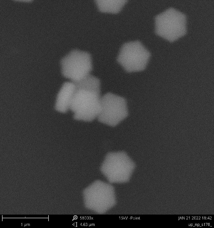
Figure 1. Photography of fluoride nanocrystals β-NaYF4:Yb3+,Tm3+, obtained on a scanning electron microscope Phenom ProX. The crystals are hexagonal, have an average diameter D≈740 nm and a height H≈260 nm.
The structural diagnostics of nanocrystals was carried out on an X-ray diffractometer Rigaku Miniflex600 (Cu, λ=1.54184 A). For this purpose, a suspension of particles in n-hexane has been applied to a glass substrate and dried at 120°C. Figure 2 shows the diffractogram of nanocrystals confirming that their crystalline lattice corresponds to the hexagonal -phase.
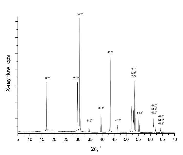
Figure 2. The diffractogram of nanocrystals β-NaYF4/Yb3+/Tm3+, obtained on the Rigaku Miniflex600 X-ray diffractometer.
Study of the size and shape of nanocrystals β-NaYF4/Yb3+/Tm3+ using luminescent optical UV microscopy
Visualization of individual nanocrystals of β-NaYF4/Yb3+/Tm3+ in light of their photoluminescence in the UV region of the spectrum is a difficult task, first of all, due to small particle sizes. According to the Rayleigh criterion, the limiting resolution R of the optical microscope is determined by formula [6]
R = 0.61 ×λ / NA, (1)
where λ is the wavelength of light on which the image of the particle is obtained, and NA is the numerical aperture of the lens used. The expression (1) defines a fundamental limitation on the resolution of the microscope, which is determined by the wavelength (at a given numerical aperture of the lens). According to formula (1), the smaller the wavelength of the PL nanophosphors, the smaller the distance scale determined by the resolution R, and the smaller the size of the nanoparticles, which shape can be allowed [7-9]. Conversely, the transition to small requires the use of special lenses which are transparent in the UV range, having small chromatic aberrations and a high numerical aperture. Poorly compensated chromatic aberrations of the objective lens result in blurring the image of the object under examination in the UV region of the spectrum even at a small spectral width Δλ of the upconversion radiation of the PL nanoparticles. Additionally, for optical microscopy near 365 nm special equipment is required: digital cameras with high photosensitivity in the UV range, narrow-band interference filters, cutoff filters for suppression of pumping radiation near 980 nm, etc.
Experiments for observing nanocrystals of β-NaYF4/Yb3+/Tm3+ were carried out using a luminescent optical microscope LUMAM IUUV-1 (LOMO, Russia) with a microphotosetting MFP 10 - U4.2, equipped with a digital UV camera SCM2020-UV-TR (EHD imaging GmBH, Germany) and an immersion mirror-lens objective 125x/1.1 (LOMO, Russia). This lens is designed to obtain images in the UV wavelength range of 250-590 nm, has an increase of 125x and a numerical aperture NA = 1.1. Excitation of luminescence of nanocrystals in the UV range was carried out by a semiconductor laser with a wavelength of ~ 980 nm. At the same time, the luminescent nanoparticles can be considered as self-luminous radiation sources. To cut the pumping light of 980 nm in front of the chamber, color glass filters UVG-2 and interference filters EO365/10 (Edmund Optics, USA) were installed. The studied nanoparticles were applied on a quartz plate with a thickness of 0.17 mm, which was installed on the microscope stage with a clean side to the objective.
Figure 3 shows a photography of the nanocrystals of β-NaYF4:Yb3+,Tm3+ in the light of the PL in the 365 ± 5 nm range. The diameter of the nanocrystals at Figure 3 is D≈630 nm, which is well suited to electron microscopy data, see Figure 1. The hexagonal shape of the particles confirming that they are in the hexagonal β-phase is clearly visible.

Figure 3. The photography of nanocrystals of β-NaYF4:Yb3+,Tm3+, obtained on an optical microscope by LUMS-IUF1 in light of the PL in the range of 365 ± 5 nm when pumped by laser radiation with a wavelength of λ≈980 nm.
In order to determine the minimum diameter of nanoparticles, the hexagonal shape of which can be distinguished on a LUBE-IUF1 microscope with a 125x/1.1 lens, nanoparticles of β-NaYF4:Yb3+, and Tm3+ of a smaller size can also be synthesized by thermal decomposition of TFA. Photographs of particles having a diameter of from 370 to 610 nm are given in Figure 4. Analysis of Figure 4 shows that with lens 125x/1.1 at wavelength 365 nm hexagonal shape of particles can be distinguished starting from diameter D≈560 nm. The shape of the particles with a smaller diameter is not possible to be determined due to the limited resolution of the optical microscope at a wavelength of 365 nm. However, the presence of luminescent nanoparticles is clearly established.
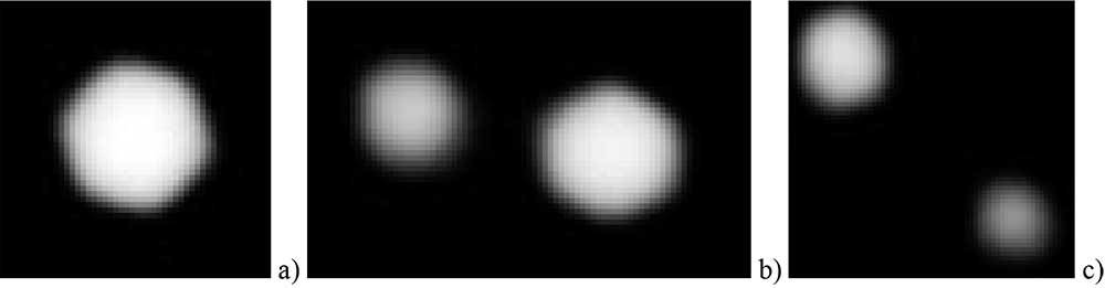
Figure 4. Photographs of nanocrystals of β-NaYF4:Yb3+,Tm3+ in light of the PL in the range of 365 ± 5 nm when pumping with laser radiation with a wavelength of λ≈980 nm. (а) The particle diameter D≈610 nm. (b) The diameter of the right particle D≈560 nm,and the left particle diameter ≈415 nm. (c) The diameter of the left particle D≈435 nm, and the right particle is ≈370 nm.
Note that the resolution, calculated by formula (1) on the photoluminescent optical microscope LUMS-IUF1 at λ=365 nm with the lens 125x/1.1 is R = 0.61 × 365 nm/1.1 = 202.4 nm. With this lens at a wavelength of λ=365 nm, a hexagonal shape of the particles can be determined starting from the diameter D≈560 nm (see Figure 4a, b), which is 2.8 times more than the resolution of R = 202.4 nm at this wavelength. Observation of nanoparticles in light of their PL at shorter wavelengths, for example, near 282 nm, where R = 0.61 × 282 nm/1.1 = 156.4 nm, presumably will allow to determine the shape of particles with a smaller diameter.
Conclusion
The size and shape of the fluoride nanocrystals NaYF4, NaLuF4, etc., doped with rare earth elements (Yb3+, Er3+, Tm3+ etc.) and having photoluminescence in the UV region of the spectrum when pumped by laser radiation with a wavelength of 980 nm, are usually examined by scanning or translucent electron microscopy. Electron microscopy has very high spatial resolution, but requires vacuity of the sample, application of metal coatings, etc. We have shown that the size and shape of the photoluminescent nanocrystals of β-NaYF4:Yb3+,Tm3+ with a diameter of 560 nm can be determined by UV optical microscopy at a wavelength of 365 nm. Unlike electron microscopy, this method is simple, fast, and makes it possible to easily visualise luminescent nanoparticles among other small objects. The use of immersion UV lenses with a larger numerical aperture and the promotion of shorter wavelengths into the direction of shorter wavelengths makes it possible to analyze the shape and size of fluoride nanocrystals with an even smaller diameter.
Acknowledgements
This work was financially supported by the Ministry of Science and Higher Education of the Russian Federation within the framework of the state task of the Federal Research Center «Crystallography and Photonics» of the Russian Academy of Sciences. The equipment of the Center for Collective Use of the FRC «Crystallography and Photonics» of the Russian Academy of Sciences was used in the work. The authors express the gratitude to G.W. Mishakov for his assistance in the preparation of experiments.
References
- H.A. Hoeppe, Recent developments in the field of inorganic phosphors, Angew. Chem., Int. Ed., 2009, 48, 3572-3582.
- D.N. Karimov, P.A. Demina, A.V. Koshelev, V.V. Rocheva, A.V. Sokovikov, A.N. Generalova, V.P. Zubov, E.V. Khaydukov, M.V. Koval’chuk, V. Ya. Panchenko, Upconversion Nanoparticles: Synthesis, Photoluminescence Properties, and Applications, Nanotechnologies in Russia, 2020, 15, 655–678.
- Mai H.-X., Zhang Y.-W., Si R., Yan Z.-G., Sun L.-D., You L.-P., Yan Ch.-H, High-Quality Sodium Rare-Earth Fluoride Nanocrystals: Controlled Synthesis and Optical Properties, J. Am. Chem. Soc., 2006, 128(19), 6426-6436.
- V.I. Sokolov, I.M. Asharchuk, I.O. Goryachuk, S.I. Molchanova, Synthesis of fluoride NaYF4/Yb/Tm, NaYF4/Yb/Er micro- and nanocrystals and their characterization by UV optical microscopy method, Fluorine Notes, 2021, 6(139), 7-8.
- V.I. Sokolov, E.N. Glazunova, I.O. Goryachuk, S.I. Molchanova, Synthesis of β-NaYF4/Yb+3/Er+3 fluoride microcrystals with in situ control of photoluminescence in up- and down-conversions, Fluorine Notes, 2023, 1(146), 3-4.
- G.S. Landsberg, Optics, Moscow, "Nauka", 1976, 928 p.
- W. Vollrath, Ultra-high-resolution DUV Microscope Optics for Semiconductor Applications, Proc. of SPIE, 2005, 5865, 58650E.
- T. Sure, T. Bauer, J. Heil, J. Wesner, DUV-Microscope objectives: technology driver that forces the production to switch from the micrometer scale to the nanometer scale, Proc. of SPIE, 2005, 5965, 59651H.
- G. Ehret, F. Pilarski, D. Bergmann, B. Bodermann, E. Buhr, A new high-aperture 193 nm microscope for the traceable dimensional characterization of micro- and nanostructures, Measurement Science and Technology, 2009, 20(8), 1-10.
ARTICLE INFO
Received 17 November2023
Accepted 11 December 2023
Available online December 2023
Recommended for publication by PhD A.A. Tyutyunov
eLIBRARY Document Number (EDN) XMSTVT

Fluorine Notes, 2023, 151, 5-6